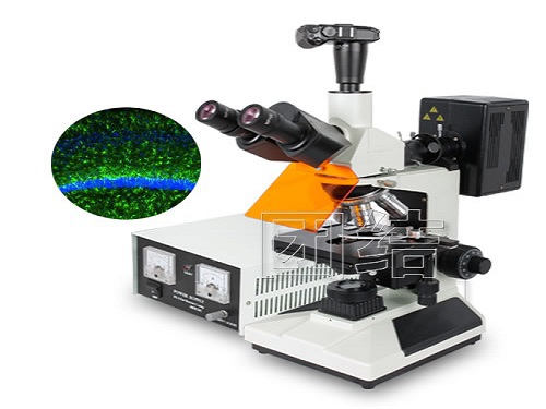
Introduction: What is a fluorescence microscope? A fluorescence microscope uses ultraviolet rays as a light source to irradiate the object to be inspected so that it fluoresces, and then observes the shape of the object and its position under a microscope. Today we will introduce specific fluorescent microscopy products and hope to help everyone.
China Cable Granulator,Wire Granulator supplier & manufacturer, offer low price, high quality Copper Wire Granulators,Cable Granulator Machine, etc.
[Conical co-rotating twin-screw cable material granulator" has many superiorities ,such as owning high yield (10 to 20 tons/day),ultra-low power consumption (lowest electricity consumption:90kwh for 1 ton of finished product), good mixing and plasticization (high-pressure and plasticization),and can add high proportion of calcium carbonate to the materials (up to 400),and also can be reproduced for 100% reclaimed material with full exhaust and high pressure .It is the best choice for all kinds of cable material granulation, such as PVC,PE,LSNH,and cross-linking,etc.
Cable Granulator,Wire Granulator,Copper Wire Granulators,Cable Granulator Machine Zhoushan Tongfa Machinery Co.,Ltd. , https://www.tongfa-machinery.com
The application of fluorescence microscopy is different from ordinary optical microscopes. Instead of observing specimens by illumination of ordinary light sources, they use certain wavelengths of light (usually ultraviolet light, blue-violet light) to excite the fluorescent substances in the specimens under the microscope to make them emit. Fluorescence, therefore, the role of the light source of the fluorescence microscope is not direct illumination, but as an energy source to stimulate the specimen's internal fluorescent substance. The reason why we can observe specimens is due to the illumination of the light source, but it is the fluorescence phenomenon exhibited by the fluorescent substances in the specimen after absorbing the excited light energy.
It can be seen that the characteristics of the fluorescence microscope are mainly that its light source can supply a large amount of excitation light in a specific wavelength range, so that the fluorescent material in the examined specimen can obtain the excitation light with the necessary intensity. At the same time, the fluorescence microscope must have a corresponding filter system.
Fluorescence microscopy is the basic tool for immunofluorescence histochemistry. It is composed of ultra-high pressure light source, filter system (including excitation and pressing filter plate), optical system and photographic system and other major components. It uses certain wavelengths of light to stimulate the specimen to emit fluorescence.
Excitation fluorescence: According to the wavelength range of light is divided into UV excitation method (using ultraviolet light method) and BV excitation method (using blue-violet light) two. The UV excitation method uses near-ultraviolet light shorter than 400 nm for excitation. This method does not have visible excitation light, so the observed fluorescence exhibits the inherent fluorescence of the dye, and it is easy to distinguish between the specific fluorescence on the specimen and the autofluorescence of the background tissue.
The BV excitation method is excited from ultraviolet to blue light centered at 404 nm and 434 nm. This method uses blue light to illuminate the specimen, so the cut-off filter of the fluorescence observation system must use a filter that can completely block blue light and sufficiently pass the required green and yellow fluorescence. Fluorescent dye for fluorescent antibody method. The maximum absorption wavelength of the excitation light and the maximum emission wavelength of fluorescence are relatively close, so the filter used in the BV excitation method must use a sharp cut filter. This method can use blue light as the excitation light, so the absorption efficiency of the fluorescent pigment is high and brighter images can be obtained. The disadvantage is that the fluorescence below 500 nm cannot be seen, and that above 500 nm makes the entire image appear yellow. In the fluorescent antibody method, the specificity is often judged by the color unique to the fluorochrome. Therefore, when the subtle specificity is discussed, the disadvantages of the above-mentioned BV excitation method often have a great influence.
In summary, the illumination of the fluorescence microscope can be considered in the following three points according to the concentrator configuration and the wavelength of the excitation light.
1 From the contrast requirements of fluorescent images, UV-excited dark field condensers provide the best illumination.
2 Considering the brightness of the image, the dark field observation efficiency of the BV excitation filter is the highest.
The characteristics of the dark field observation of the 3UV excitation filter and the dark field condenser illumination of the BV excitation can be regarded as between these two illumination methods, but the dark field of the former filter is stronger and the display image is brighter. The contrast is small; in the latter, because the nature of the dark field light path is preserved, the display image is darker and the contrast is improved. When a fluorescence microscope is actually used, observation should be performed using the illumination method best suited to the requirements of the specimen.
It must be pointed out that, even with the best-contrast UV-excited dark-field condenser illumination, part of the UV-excited light refracted or scattered by the specimen enters the objective lens, resulting in excitation of the Canadian adhesive bonding surface and optical glass in the optical system. Autofluorescence can make the reaction worse. Therefore, UV absorption filters must be used as cut-off filters in front of the eyepieces.
Application of fluorescence microscope
